

 By Dr. Stefanie Novakowski, Kastrup Lab
Dr. Karen Cheung is a professor at UBC in the Department of Electrical & Computer Engineering and the new School of Biomedical Engineering. She is also the Director of the Graduate Biomedical Engineering Program. As one of the newest members of the CBR, having joined in June, Dr. Cheung’s recruitment represents exciting new directions for blood research at the CBR and its translation to the clinic.
Dr. Karen Cheung
Can you tell me a bit about your research background?
I studied bioengineering as an undergraduate student, followed by a PhD also in bioengineering at the University of California, Berkley. In high school I liked both sides, the biology side and the engineering side, and I grew up in California, where bioengineering was available as an undergraduate degree program.
How have the opportunities for high school or undergraduate students interested in bioengineering changed since you started your career?
When I was an undergraduate student, the program was not structured as well as programs are now. Instead of just taking some engineering courses and some biology courses, programs now are designed to help students make connections between the molecular level, cellular level, tissue level, and functional level, considering both the engineering and biological components.
What are some of the research projects your lab is focused on now?
I am involved in several projects including, for example, organ-on-a-chip and cellular printing. We have a tumour-on-a-chip wherein we culture spheroids, which are models of tumours. We make them three-dimensional, so they actually model multi-cellular structures, and include not just cells found within the tumour, but also extracellular components, like the proteins important for cell attachment. We use hydrogel beads to grow the cells, and this allows us to study the effect of hypoxia on the cells in a dynamic matter, as these conditions change over time in the body. With microscopy, we study in real-time, what’s happening to the cells. We are also collaborating with colleagues at St. Paul’s Hospital to create an airway-on-a-chip. There, we are modeling a very specific part of the lung and studying the effects of fibrosis in chronic obstructive pulmonary disease and asthma.
In terms of cellular printing, our work focuses on flow hydrodynamics in inkjet printing. When you are printing cells, the cell diameter is very close to the size scale of the inkjet nozzles. Our research was the first to see what’s happening inside the nozzle, close to the cell, using high-speed imaging. This lets us study how different properties of the ink affect the cells during the printing process.
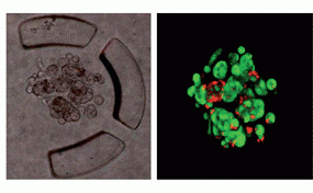
Tumour-on-a-chip: Breast tumor cells growing and forming multicellular spheroids, encapsulated within gel-like droplets. Individual cells are labelled in green (right). Adapted from Yu L, Chen MC, Cheung KC. Lab Chip. 2010;10(18):2424-32.
Is the focus of your lab on the biology or building the tools and models for studying the biology?
Ideally, it’s both. We’re not just building bridges for other people, although this really drives us in terms of what we build, because it would be nonsensical to make something and tell other researchers it’s what they need. The hypoxia study arose from conversations with researchers at the BC Cancer Center, who knew it was important to study the hypoxic conditions in a dynamic setting. This is what drove us to spend time developing this technology. To do this, I try to build a group of students who come in from different backgrounds and then complement each other. That way we use the natural strengths of each trainee, depending on their background; but there are still opportunities for them to work together and learn from each other.
How do you think your research fits in with the CBR, in terms of their overall goal and their ongoing projects?
While we’ve been making tumor models with our technology, we can easily build different types of models with these tools that would be relevant to other researchers at the CBR. For the inkjet project, in addition to tissue engineering, we are trying to dispense single cells. If we are able to rapidly dispense single cells in a high-throughput way, we can use this for single cell studies, which should lead to additional collaborative opportunities.
What excites you the most about your work? What work are you most proud of?
What’s most exciting about research in general is exploring things that have not been solved before. In my lab, we are building technologies that enable scientists to do things they can’t do yet. Once we have achieved that, we would move forward to working with partners to make fundamental discoveries. It’s exciting to create these new technologies, and I’m very proud of my trainees who have the perseverance and patience to stick through it. There are always lots of challenges, but they overcome them, and I am very proud of that.
If you had any advice for trainees, what would it be?
Be open to investigating new things. One of my challenges that I did not foresee in the organ-on-a-chip project, was how difficult it would be to image the cells. My students were not afraid to explore new microscopy techniques, regardless of what their academic background was.