


By Dr. Shawna Stanwood, Jefferies Lab alum
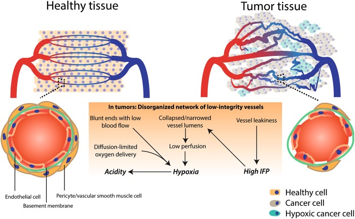
Figure 1. The blood vessels of tumours. IFP refers to interstitial fluid pressure. This image has been reproduced from the publication by Schaaf et al. without modification and is being used with permission under a Creative Commons Attribution 4.0 International License. Link: http://creativecommons.org/licenses/by/4.0/
“Toto, I’ve a feeling we’re not in Kansas anymore.” – The Wizard of Oz 1
While Dorothy was certainly not talking about tumour research, one thing is for sure when it comes to cell culture: we’re not in 2D anymore.
The words “cancer” and “hypoxia” frequently come up in the same conversation (in some cases, literally)2. Defined by the National Cancer Institute as a “condition in which there is a decrease in the oxygen supply to a tissue,” hypoxia can be exhibited by certain cells within a tumour due to problematic blood vessels (Figure 1) 3, 4. Cell culture that reflects the 3D nature of tumours can help researchers learn more about not only how tumours behave within the framework of hypoxia, but also how they react to treatment. This latter point is of particular interest given that hypoxia seems to hamper various kinds of therapy5.
In their paper published in Scientific Reports, Dr. Karen Cheung (Centre for Blood Research) and researchers in the Department of Electrical and Computer Engineering at UBC conveyed their findings from experiments conducted using 3D tumour spheroids6. To construct the spheroids, the team grew breast tumour cells inside beads (Figure 2). Beads bearing spheroids were then used in “on-chip studies” involving methods that enabled the control of oxygenation at the microfluidic level (see Figure 2C for scale).
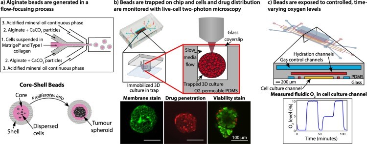
Figure 2. Tumour spheroids and on-chip experiments. This image has been reproduced from the publication by Grist et al. without modification and is being used with permission under a Creative Commons Attribution 4.0 International License. Link: http://creativecommons.org/licenses/by/4.0/
The concentration of O2 that is available can differ depending on the context. Although O2 usually makes up 18.6% of the atmospheric pressure within an incubator used for cell culture, the physiological percentage when referring to tissues of the periphery (“physoxia”) is actually much lower (and even lower for various forms of hypoxia) 5, 7. By bringing into play different percentages of O2 in a cyclic manner, first author Dr. Samantha Grist and colleagues found that an absence of O2 (i.e. 0%) resulted in swelling of the spheroids, whereas 3% and 10% O2 prompted shrinking. The remarkable back-and-forth nature of this finding can be seen in Movie S1 of the Supplementary Materials.
The researchers sought to ascribe the swelling to either “cytoplasmic swelling” (which refers to individual cells expanding) or spheroid “disaggregation” (which refers to the cells not touching one another anymore). Breast tumour spheroids underwent harsh hypoxia (i.e. 0% O2) followed by “normoxia” (i.e. 20% O2) or vice versa. The authors found that spaces between cells did not seem to develop. Furthermore, when average cell size was explored, it was found that cells subjected to 0% O2 became enlarged more quickly compared to cells subjected to 20% O2. These findings, therefore, seemed to support the first hypothesis, namely the one based on swelling of the cells themselves.
The authors also delved into how oxygenation shapes the uptake of drugs used to fight cancer, specifically doxorubicin. This drug can be used (either by itself or in combination with other drugs) to combat breast cancer via chemotherapy 8. Spheroids experienced 0% O2, 20% O2, or O2 cycling back and forth between these two percentages while undergoing treatment with the drug for 72 hours. Greater build-up of doxorubicin (indicated by its inherent fluorescence) was found when the oxygen followed a cyclic pattern. Moreover, the research group found that certain cells in the spheroid displayed stronger fluorescence than that of neighbouring cells. Expression of P-glycoprotein, migration of cells, and pH were put forward as hypothesized explanations for this non-uniform trait.
The outcomes of this study reveal what happens to tumour spheroids in hypoxic circumstances. The authors also pointed out that future research, such as an effort to learn more about the mechanism, may uncover more about cancer, both in terms of its treatment and its progression.
References:
1 What Is the Greatest Movie Quote of All Time? The Atlantic [Internet]. 2019 Sep [cited 2020 Mar 26];The Big Question:[about 5 p.]. Available from: theatlantic.com/magazine/archive/2019/09/q-what-is-the-greatest-movie-quote-of-all-time/594748/.
2 Pancholi, NJ. Hypoxia and the Lethal Cancer Phenotype: A Conversation with Nobel Laureate Gregg Semenza. American Association of Cancer Research. June 5, 2020. https://www.aacr.org/blog/2020/06/05/hypoxia-and-the-lethal-cancer-phenotype-a-conversation-with-nobel-laureate-gregg-semenza/
3 National Cancer Institute. NCI Search Results – Results for: Hypoxia. National Cancer Institute. https://www.cancer.gov/search/results?swKeyword=Hypoxia
4 Schaaf, M.B., Garg, A.D. & Agostinis, P. Defining the role of the tumor vasculature in antitumor immunity and immunotherapy. Cell Death Dis 9, 115 (2018). https://doi.org/10.1038/s41419-017-0061-0
5 Wenger RH, Kurtcuoglu V, Scholz CC, Marti HH, Hoogewijs D. Frequently asked questions in hypoxia research. Hypoxia (Auckl). 2015 Sep;3:35-43. https://doi.org/10.2147/HP.S92198
6 Grist SM, Nasseri SS, Laplatine L, Schmok JC, Yao D, Hua J, et al. Long-term monitoring in a microfluidic system to study tumour spheroid response to chronic and cycling hypoxia. Sci Rep. 2019 Nov;9(1):17782. https://doi.org/10.1038/s41598-019-54001-8
7 McKeown SR. Defining normoxia, physoxia and hypoxia in tumours – implications for treatment response. Br J Radiol. 2014 Mar;87(1035):20130676. https://doi.org/10.1259/bjr.20130676
8 Canadian Cancer Society. Chemotherapy for breast cancer. Canadian Cancer Society. https://www.cancer.ca/en/cancer-information/cancer-type/breast/treatment/chemotherapy/?region=on
9 Trédan O, Galmarini CM, Patel K, Tannock IF. Drug resistance and the solid tumor microenvironment. J Natl Cancer Inst. 2007 Oct;99(19):1441-54. https://doi.org/10.1093/jnci/djm135