


By Rhonda Thygesen, MSc Student, Foster Lab
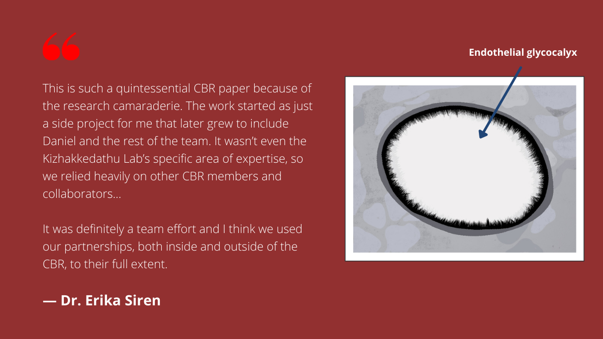
Sitting at the interface between blood flow and the endothelial cells that line our blood vessels are glycocalyces, brush-like structures formed mostly of carbohydrates that protect the tissue. The glycocalyx acts in physiological responses such as innate immunity, inflammation, and coagulation by structural change. In order to explore these biological processes, a researcher must be able to grow proper endothelial cells with accurate glycocalyces.
Recently, the Kizhakkedathu Lab at the Centre for Blood Research (CBR) and their collaborators developed an improved method for studying glycocalyces, one that can mimic the complex environments of an organism’s vascular system, by using microfluidic chip technology.1 Former PhD Dr. Erika Siren and PhD candidate Daniel Luo shared their reflections about the study, future directions for this work, and the collaborative efforts of the CBR that made this research possible.
ES: Dr. Erika Siren, DL: Daniel Luo

Daniel Luo
What was your study about?
ES: One of the biggest issues with studying the glycocalyx in vitro is that it doesn’t grow well in static cell culture. It’s important that the glycocalyx grows properly in length and thickness, as that will affect its physiological properties in experiments.
In the body, the glycocalyx actually grows under the shear stress of blood flow. With that in mind, we wanted to develop a device that would recreate the fluid shear stress and better mimic these natural conditions. That’s where the microchip idea came from. Ultimately, our goal is to make a better, more physiologically relevant model for researchers studying the endothelial glycocalyx.
DL: This methods paper demonstrates that our microchip technology works, but also reveals that the environment in which a glycocalyx grows – in static cell culture or under shear stress – affects subsequent experiments. Our paper attempts to reveal this grey area.

Dr. Erika Siren
This project involved many collaborators, from the CBR and beyond. How did the expertise of different researchers make this project a success?
ES: This is such a quintessential CBR paper because of the research camaraderie. The work started as just a side project for me that later grew to include Daniel and the rest of the team. It wasn’t even the Kizhakkedathu Lab’s specific area of expertise, so we relied heavily on other CBR members and collaborators.
I remember walking down the street to Dr. Karen Cheung’s lab to ask a few questions when we first started making the microchip. It was awesome, because their lab had all the equipment, facilities, and expertise – they helped with the microchip’s development, from understanding parameters to teaching us how to seed the cells into the chip.
When I needed to perform computational simulations and ensure that the forces in the chip were accurate, we partnered with Dr. Jordan Mackenzie and Dr. Mark Martinez across the street. The Conway Lab, which is right next to our lab bench in the Life Sciences Centre, was also an invaluable resource. Dr. Ed Conway really helped put the impact of our methodology into context, thanks to his strong background in immune regulation, coagulation, and complement.
It was definitely a team effort and I think we used our partnerships, both inside and outside of the CBR, to their full extent.
What made you the most excited about this research?
ES: There were many times when I encountered barriers that I couldn’t overcome alone. It was amazing to be able to literally reach out across the hall, street, or bench to someone who had the resources to help. That’s what scientific research is – resourcing the right people and expertise around you to solve problems.
“That’s what scientific research is – resourcing the right people and expertise around you to solve problems.”
– Dr. Erika Siren
What are the future directions for this project? How might other researchers use this method?
DL: We’re starting to see a lot more interest from outside of our own circle and from collaborators who are interested in working with us on similar projects involving the glycocalyx. Because of this, our paper’s findings are frequently referenced. Having this paper published is helpful for showing that there is a big difference between standard static cell culture and our current in vitro platform.
ES: As Daniel mentioned, microchips with cell cultures can act as a middle step between static cell culture studies and in vivo studies, providing useful data that can help optimize conditions for later animal studies. In doing so, this technology can reduce the expenses as well as time associated with animal studies.
Microchip technology is also extremely tunable and adjustable, and can be tailored to answer different research questions. For example, a scientist could use this technology to grow and study glycocalyces that are specific to certain organ systems, or glycocalyces specific to certain animal models. They could also try using different cell types and co-cultures on the microchip, or add additional cell layers to further replicate the blood vessel. All in all, it’s a very versatile method with lots of potential applications.
References
1 Siren EMJ, Luo HD, Bajaj S, et al. An improved in vitro model for studying the structural and functional properties of the endothelial glycocalyx in arteries, capillaries and veins. The FASEB Journal (2021) 35(6) doi:10.1096/fj.201802376rrrr