

 By Soroush Nasseri, PhD Student, Cheung Lab
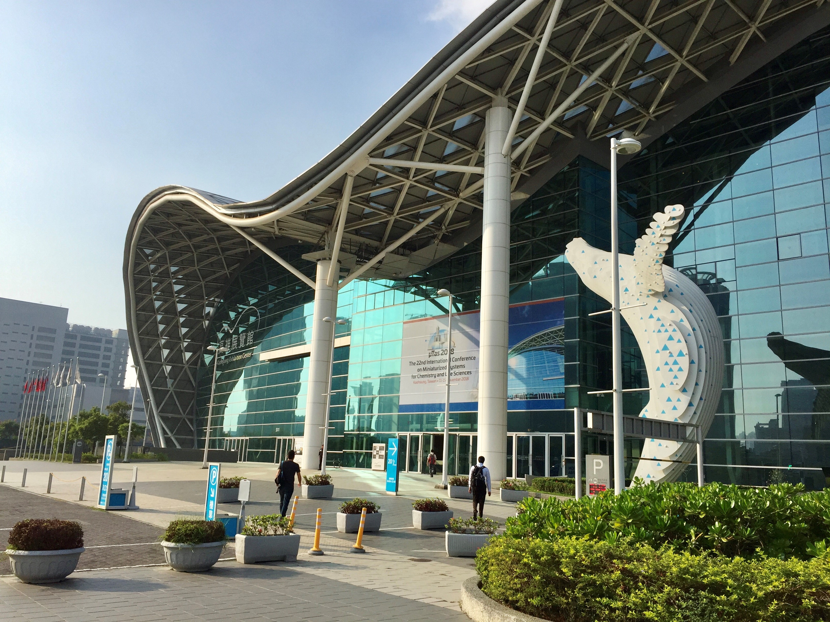 Recently, I had the opportunity to attend The 22nd International Conference on Miniaturized Systems for Chemistry and Life Sciences (µTAS 2018) hosted by Taiwan’s National Tsing Hua University in Kaohsiung. The conference, organized every year by The Chemical and Biological Microsystems Society and rotating among Asian, European and North American venues, is one of the most prestigious scientific conferences in the field of microfluidics. The conference places great emphasis on the biological applications of microfluidics and lab-on-a-chip systems, looking for more efficient ways to answer pressing questions in life sciences, and providing new vehicles to explore uncharted territories.
µTAS 2018 featured more than 2400 abstracts (oral presentations and posters) plus industrial exhibitions and workshops. While µTAS included many seemingly unrelated sessions, these could be categorized into three main themes: microfluidic technology per se, application of microfluidics in clinical diagnosis, and application of microfluidics in chemical and biological research. Some topics that caught my attention were several attempts at organ-on-a-chip devices for research or personal medicine, and microfluidic devices for diagnosis of diseases more rapidly or more economically.
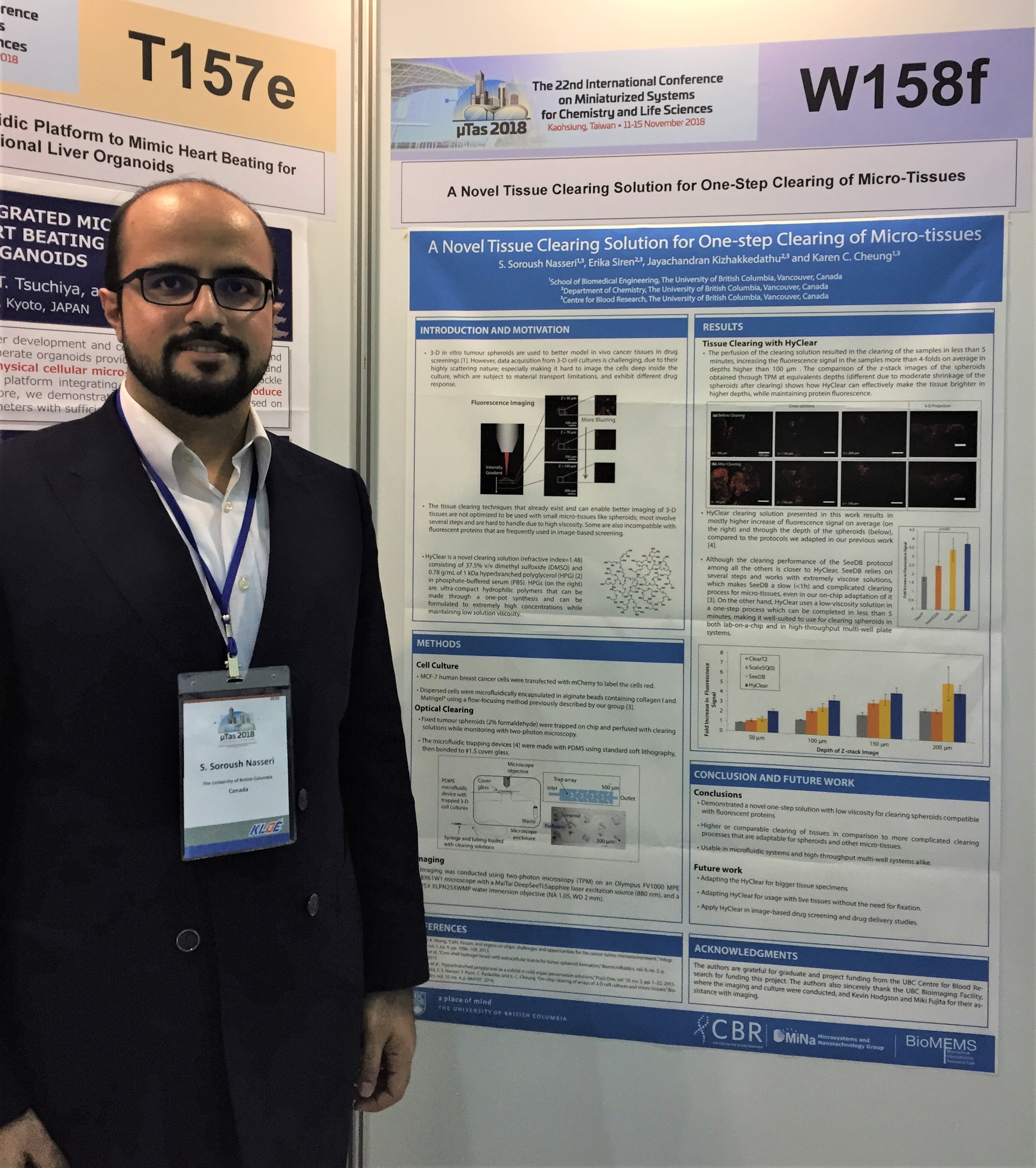 I presented a poster of our work in Dr. Karen Cheung’s lab in collaboration with Dr. Kizhakkedathu. This work concerns tissue clearing for better microscopy of 3D microtissues. 3D microtissues are novel models for mimicking in vivo tissues, standing between traditional cell cultures and animal models. One of the benefits 3D microtissues provide is the ability to readily image them with confocal microscopy or other techniques. However, physical properties of tissues prevent imaging deep into the tissue; as we go deeper, fluorescent signals get blurry and eventually are lost. This problem is traditionally addressed with a laborious embedding and sectioning process. Dr. Cheung’s lab has eliminated the need for this process by using the properties of hyperbranched polyglycerols (HPG) to enable the 3D microscopy of whole microtissues. This will allow us to derive more informative data from all parts of the 3D microtissues, and will support the development of a rapid and high-throughput image-based drug screening platform. I very much enjoyed talking to the people about my work, and conceived new ideas on how to make progress in the project based on comments I received from the people who would benefit from using our improved clearing techniques in their research.
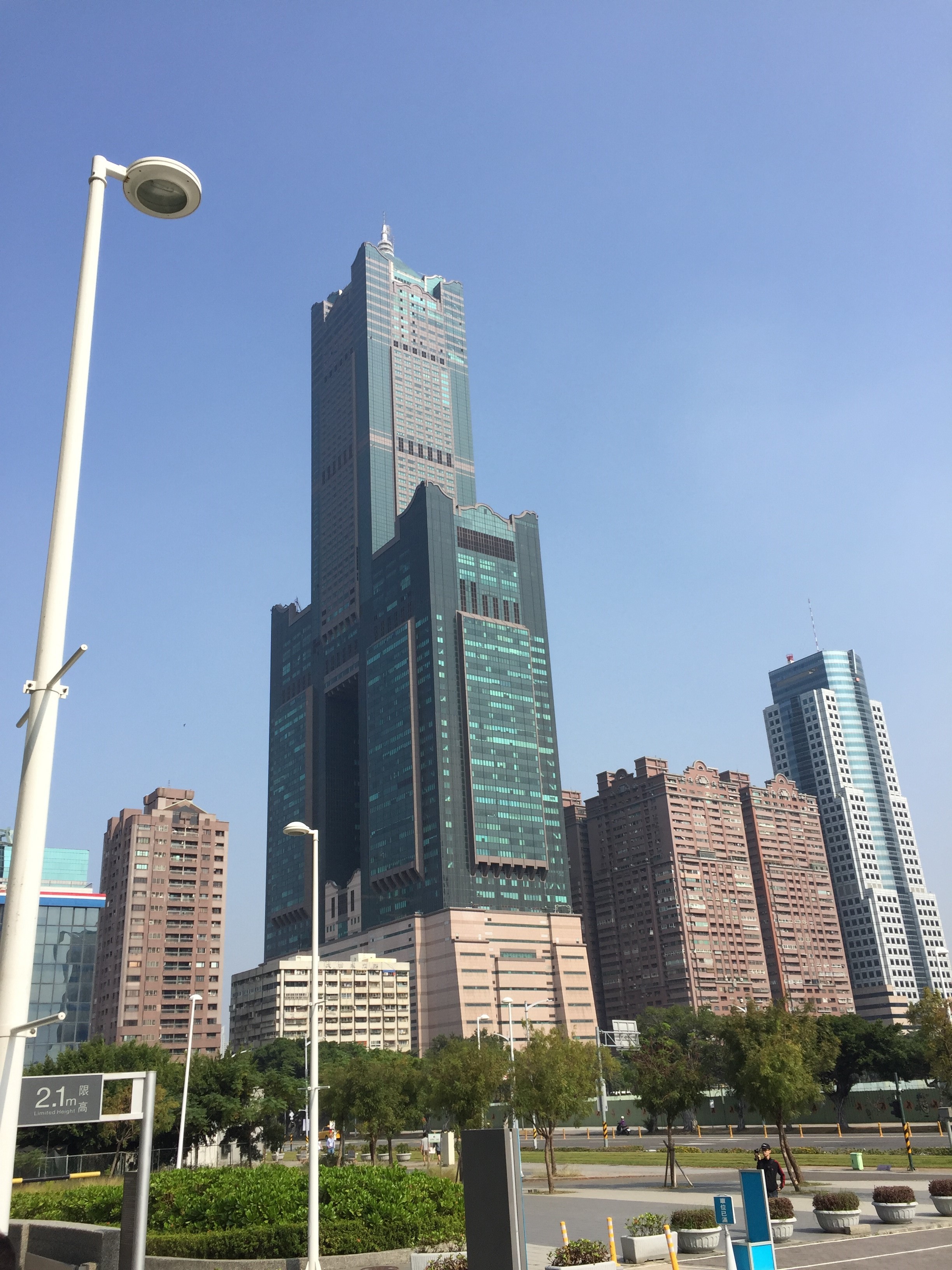 Attending a conference also provides opportunities for fun! Although the talks and posters were too good to miss, I still found some time in the free hours and after the conference to enjoy the beauty of Taiwan. The conference was held in Kaohsiung, one of the more populous cities of Taiwan in the south of the island. At the time when Vancouver weather had everyone shivering, Kaohsiung was easily keeping it up above 25 degrees! It is a beautiful city with much to explore: natural beauties, cultural attractions, and lively night markets. I also stayed in Taipei for a couple of days and visited the National Palace Museum, which contains many artefacts of Chinese civilization. The highly efficient railways of Taiwan also made it easy for me to travel to Hualien for a day and explore the famous Taroko national park, with its wonderful gorge and many historical monuments. I will long remember Taiwan for its lively vibe, kind and hospitable people, great food and amazing natural beauty, and if the opportunity arises, would definitely recommend it as a country to visit!