

 By Fennie Easton van der Graaf, Undergraduate Student, Jefferies Lab
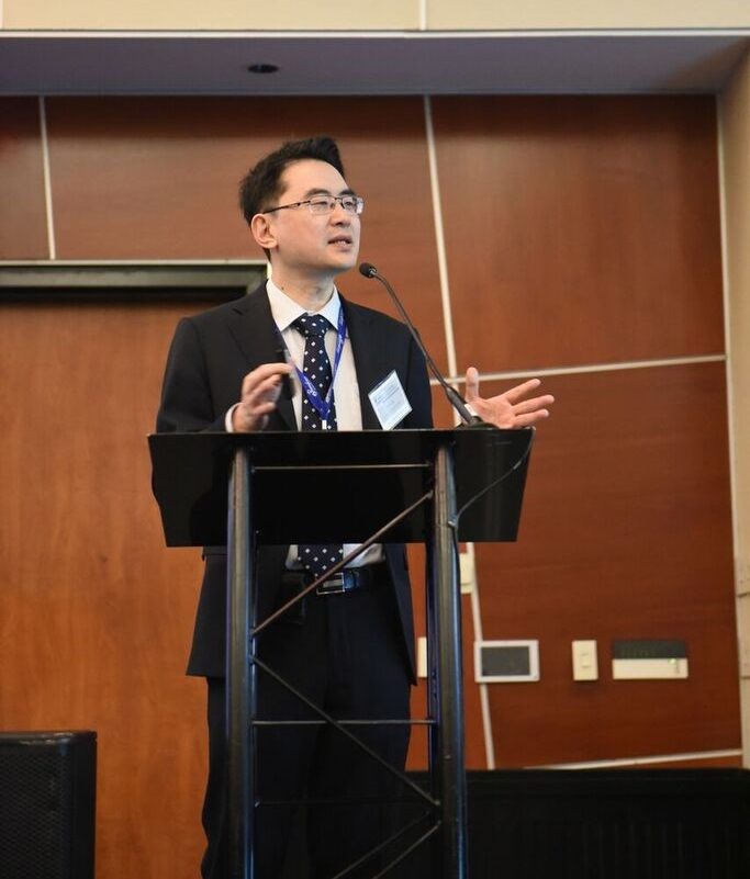
Dr. Don Sin introducing the conference.
Searching for collaborations in hospitals, industry, or academia? Passionate about improving care for patients with airway disease? The ‘Creation of a Pan-Canadian Airway Network Conference’ provided the perfect opportunity to forge connections.
Facilitated by COPD expert, Dr. Don Sin, and Program Manager, Dr. Tony Yang, the November 5th Pan-Canadian Airway Network Conference cleverly addressed medical goals to combat Chronic Obstructive Pulmonary Disease (COPD) from an array of scientific angles. The scope of this conference united a community of clinicians, biomedical engineers, geneticists, industry representatives, and health economists, all striving to bring ‘exceptional care through exceptional science’ for the over 2.5 million Canadians suffering from COPD.1
The conference consisted of four distinct sessions: Bioimaging, Biomedical Engineering, Genetics/Genomics, and Clinical Translation. The Chair of each session moderated 2-3 presentations from distinguished local and national investigators, followed by their engagement in a panel discussion. The audience was invited to ask questions either online or through a microphone, generating an intellectually stimulating but also relaxed dialogue.
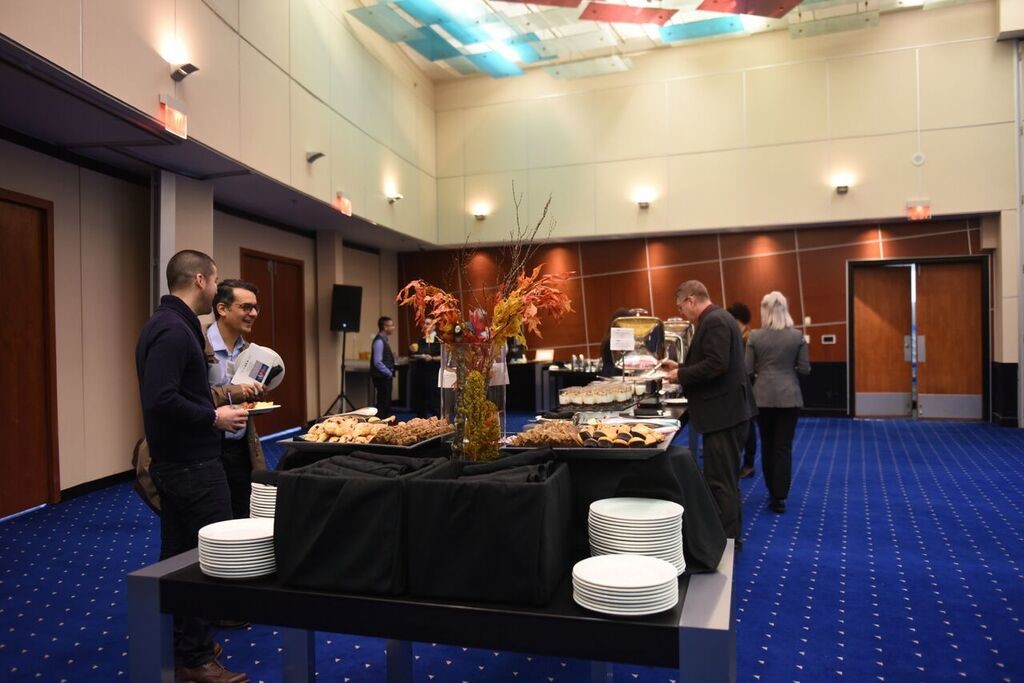
Networking and Lunch.
The chair of the UBC Department of Radiology Dr. Jonathon Leipsic set the stage for Dr. Grace Parraga (Robarts Research Institute) and Dr. Jonathan Rayment (B.C. Children’s Hospital Research Institute). Dr. Parraga evaluated the use of ‘Hyperpolarized gas Magnetic Resonance Imaging’ as a fast diagnostic tool used to convey information about COPD severity. Despite being an expensive technique, it does overcome conventional challenges of using a spirometer to measure lung function.2 Dr. Rayment expanded on this technique’s significance as it can be used to calculate ventilation defect percent (VDP)3, which is a sensitive indicator of lung disease in cystic fibrosis. His work as a pediatrician opened the suggestion to focus research on the origin of COPD and also highlighted the challenges of diagnosing COPD in young children.
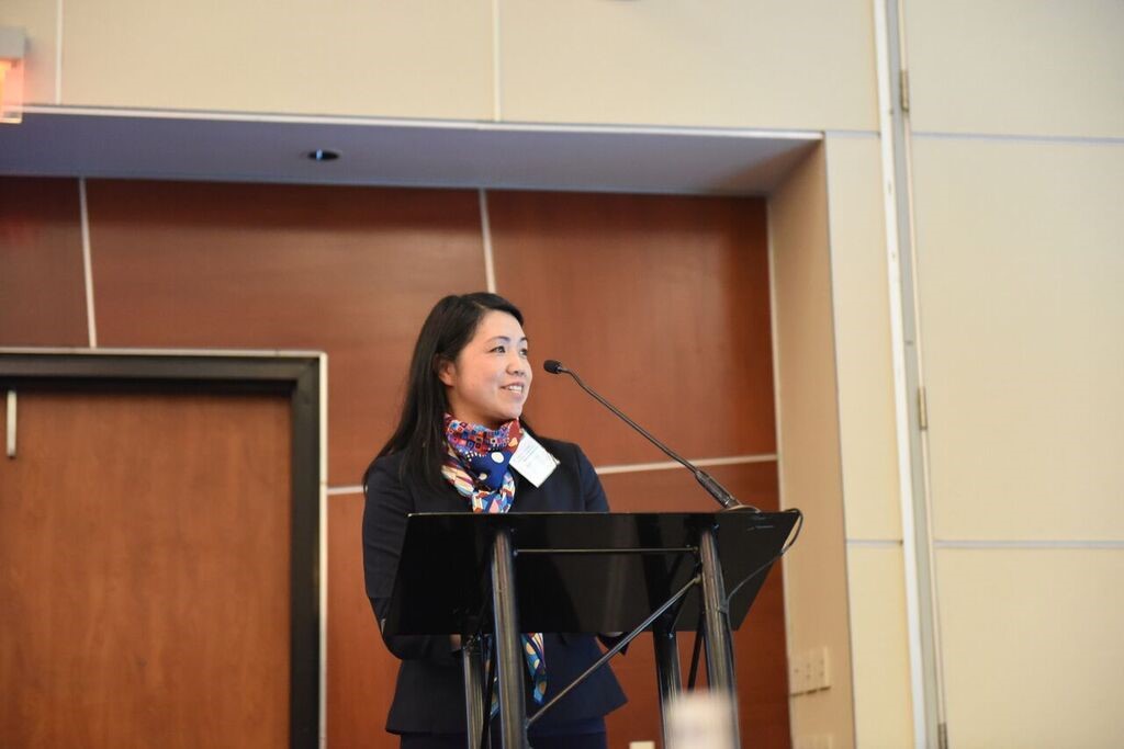
Dr. Karen Cheung introducing the Biomedical Engineering section.
Dr. Karen Cheung (a Director of the UBC School of Biomedical Engineering and PI in the Centre for Blood Research) introduced Dr. Edmond Young (University of Toronto) and Dr. Jeremy Hirota (McMaster University). Dr. Young introduced the applications for organ-on-a-chip, the method of growing lung and other tissues in hydrogel or other matrices within the chip, and how conditions can be controlled, such as ash particles passing by smooth muscle and epithelial cells, to study slight changes in environment. Dr. Hirota emphasized the significance of using flow systems in microfluidic devices to study tissues in a chip, and how he partners with Aspect Biosystems for 3D tissue printing. Such devices could be the next alternative to animal models, presenting a more affordable, ethical method to study human tissue with greater experimental control.
Through a very professional and candid atmosphere, the experience of this conference was intellectually exhilarating. I can’t tell if it was because of absorbing so much information or just eating too much of the delicious lunch (see pictures; all photo credits go to Jasemine Yang), but I really grew as a researcher throughout the course of this event. This experience truly inspired a sense of a greater cause and the ability to make a greater impact because of the support and collaboration promoted between a vast array of scientists. Be sure to attend future sessions of this conference which are likely to be held every 2 years, and contact Dr. Tony Yang for any questions: Tony.Yang@hli.ubc.ca
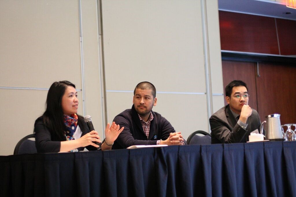
Panel Discussion: Dr. Karen Cheung (left), Dr. Jeremy Hirota (centre), and Dr. Edmond Young (right).
- https://www150.statcan.gc.ca/n1/pub/82-003-x/2014003/article/11908-eng.htm
- Fain, S., Schiebler, M. L., Mccormack, D. G., & Parraga, G. (2010). Imaging of lung function using hyperpolarized helium-3 magnetic resonance imaging: Review of current and emerging translational methods and applications. Journal of Magnetic Resonance Imaging,32(6), 1398-1408. doi:10.1002/jmri.22375
- Santyr,G., Kanhere,N., Morgado, F., Rayment, J., Ratjen, F., Couch, M.J. (2018). Hyperpolarized Gas Magnetic Resonance Imaging of Pediatric Cystic Fibrosis Lung Disease, Journal of Academic Radiology. DOI: 10.1016/j.acra.2018.04.024