

For this round of the CBR Cover Art Contest, members submitted both illustrations and images to share their labwork. The winning image, featured on cover of our November 2022 CBR Magazine, was submitted by Nooshin Safikhan of the Conway Lab.
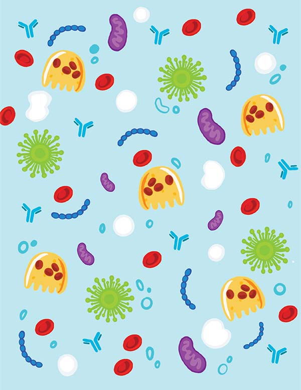
“The joy of studying things we don’t see”
Jimin Jung
MSc Student, Brömme Lab
“I love research and studying things we don’t see with our naked eyes. Here, I drew some things we might not see in person (without microscopes), but we know they exist through the wonder of research: osteoclasts, red and white blood cells, viruses, antibodies and mitochondria.”
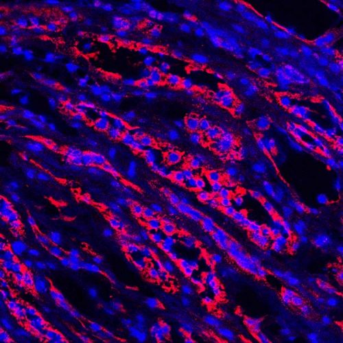
“To B Human”
Matthew Drayton
Research Assistant, Kizhakkedathu Lab
“A kidney section from a blood type B donor. Cell nuclei are stained blue, and B antigens are stained red.”
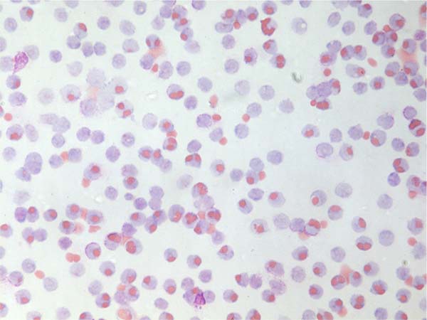
“Monocytes in Action”
Matthew Drayton
Research Assistant, Kizhakkedathu Lab
“A monocyte monolayer assay is often used to determine the compatibility of red blood cells for blood transfusion. In this case, A-type red blood cells are opsonized with anti-A antibodies from an O-type donor’s serum; the monocytes recognize the antibodies coating the red blood cells and engulfs them.”
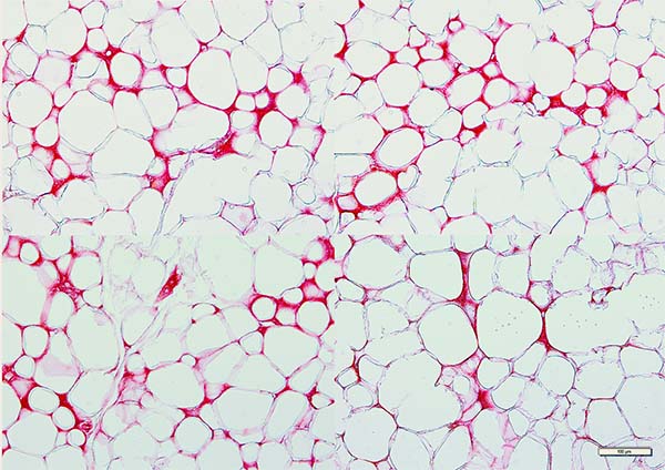
“Picrosirius red staining of collagens in fat tissue”
Nooshin Safikhan
Research Assistant, Conway Lab
“Picrosirius red staining of collagens show fibrosis (ECM deposition) in white adipose tissue of wild type CD248 after feeding with a High Fat diet (HFD) for several weeks.”
Thank you to everyone who submitted an entry and shared their science!