For this round of the CBR Cover Art Contest, we saw a creative mix of entries that provided a sneak peek into the work of different labs! The winning image, featured on cover of our April 2022 CBR Magazine, was submitted by Dr. Manoj Paul of the Kim Lab.
"Platelets at work!"Stages of transformation, from a small platelet into formidable defense structures against blood loss, shown in a single view. |
|
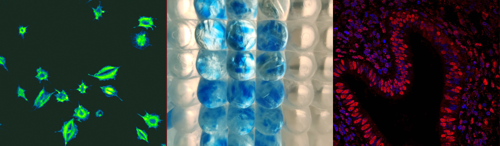
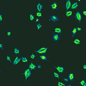
What's the story behind this image?"Platelets play a quintessential role during injury. In their inactive state, platelets look like small "plates" or discs. However, when a blood vessel is damaged, platelets will transform into their active state and undergo a dynamic change in their physical structure, quickly growing arm-like structures that help with forming blood clots. My work tries to understand how platelets function, and how a specific cytoskeletal protein called gelsolin can influence them. In my lab, I came across this single frame that showed platelets at different stages of their activation status. It is intriguing how a small, inactive platelet — which you can see at the bottom centre of the image — has the ability to rapidly transform itself into a sticky, life-saving tool with cellular "limbs"!" |
|

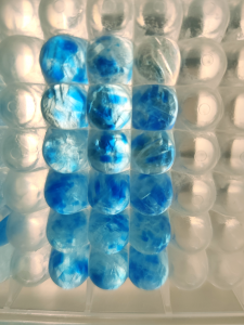
"Sapphire in-gel digestion of proteins"Blood treated with lipid nanoparticles containing protein corona is separated in a gel, before being cut into small gel pieces for downstream proteomics analysis. |
|
What does this image show? How does it relate to your research?"This image shows gel pieces in a well plate, viewed from the bottom. It is part of a proteomics procedure known as in-gel digestion, for identifying and quantifying proteins that are obtained by running gel electrophoresis. These gel pieces are blue because they were stained with Coomassie stain, a blue dye used to visualize proteins. I enjoyed looking at my proteins as they were arranged in these gel pieces, and their color reminded me of sapphire jewelry. My research focus on Corona proteins (proteins that bind to lipid nanoparticles) forming in blood cells or plasma. This investigation is part of a Mitacs project between Acuitas Therapeutics Inc, a world leader in the development of lipid nanoparticles for mRNA drugs and vaccines, and the Foster Lab at UBC, which identifies candidate proteins and discovers mechanisms-of-action through mass spectrometric analysis of proteins." |
 |

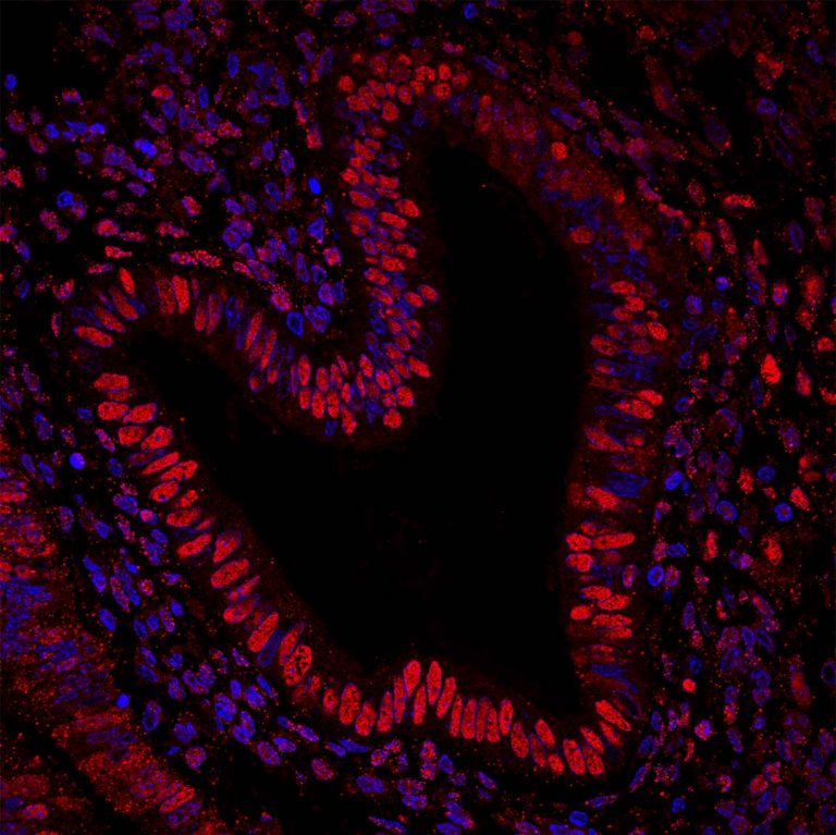
"Staining of HMGB1 in human endometrium"
|
|