

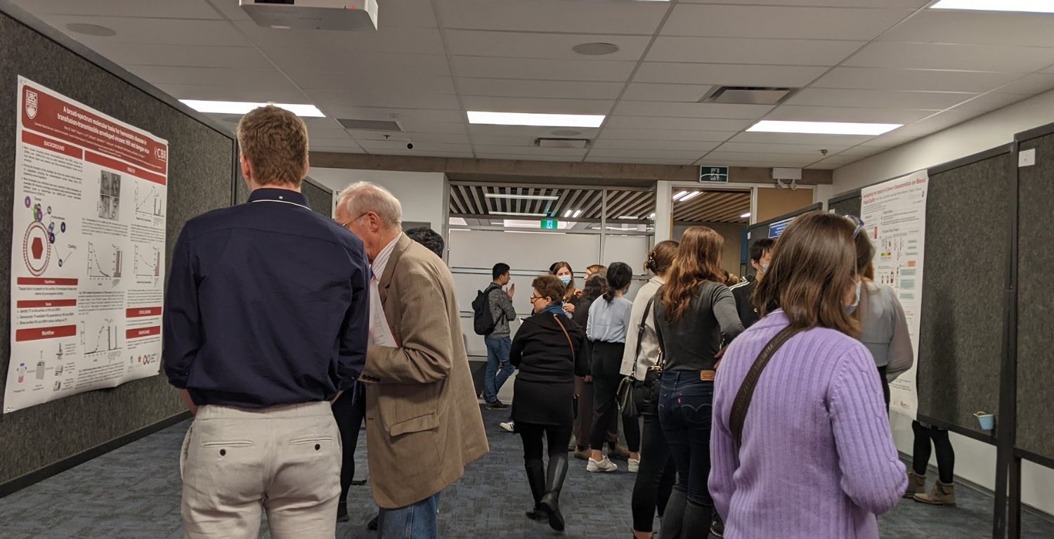
This gallery showcases poster presentations and oral talks that were presented at the 16th Earl W. Davie Symposium on November 22, 2022, at UBC Robson Square. These individuals are undergraduate students, graduate trainees, postdoctoral fellows, clinical fellows, and research associates.
- Oral Talks: During the Symposium, the audience voted for their favourite oral talk, and the winner received the People’s Choice Award: Colton Strong, PhD Candidate from the Devine & Kastrup Labs.
- Posters: During the Symposium, the posters were judged by poster judges, and the winner received the Best Poster Presentation Award: Henry West, MSc Student from the Pryzdial Lab.
If you have questions about Earl W. Davie Symposium 2022 posters, oral talks or voting for oral talk presenters please contact the CBR’s Education Program Manager, Dr. Parvin Bolourani (parvin@mail.ubc.ca).
Oral TalksThe recipient of the People’s Choice Award was Colton Strong, PhD Candidate from the Devine & Kastrup Labs.
Authors: Ahmed Kabil*, Kelly McNagny Biomedical Research Centre, The University of British Columbia, Vancouver, Canada PI: Kelly McNagny Role: Ph.D. Student A novel transgenic mouse model for Innate lymphoid cell lineage tracing and fate mapping Innate lymphoid cells (ILCs) are frontline immune-modulatory cells involved in the early stages of host defense and maintenance of tissue homeostasis, particularly at mucosal surfaces such as the small intestine, lung, and skin. The ILC family is classified into five major groups: NK, ILC1s, ILC2s, ILC3s, and LTi, based on their developmental trajectories and functional characteristics. Their significance in specific immune responses and diseases is only beginning to be understood, and the mechanisms by which ILCs regulate inflammatory conditions still remains elusive. To pinpoint the functional role of ILC2 in vivo, we have now developed Il17rbeGFP-CreERT2 transgenic mice and conducted several experiments to show that they express tamoxifen-inducible Cre-ERT2 and GFP under the control of the endogenous Il17rb promoter/enhancer. This unique mouse model enables selective lineage tracing of Il17rbexpressing ILC2s and ILC2 progenitors across all anatomical locations. The fact that Il17rb selectively marks ILC2s in vivo demonstrates that Il17rb-eGFP-CreERT2 mice are an attractive tool for studying ILC2 development, migration, and plasticity in homeostatic conditions and inflammatory diseases.
1Centre for Blood Research, 2Department of Pathology and Laboratory Medicine PI: Ed Pryzdial Role: MSc Student Modified clotting factor X: Toward a safer clot buster Heart attack and stroke are the leading causes of death worldwide and usually involve a deadly blood clot, which blocks the flow of blood. These blood clots are currently being treated by a recombinant (r) form of tissue plasminogen activator (tPA), the enzyme that would normally initiate the process of clot-busting and restore blood flow. However, when used as a medicine, rtPA must be given at very high doses, and can cause life-threatening bleeding because of its enzyme function. The Pryzdial lab shown that clotting factor (F) X can be modified to become a non-enzymatic tPA accelerator that enhances clot busting. In pre-clinical studies, this new compound called Xai-K was shown to be just as effective as rtPA at restoring blood flow in benchtop and animal models of blood clots, while being a safer alternative. Unfortunately, Xai-K has many production steps and is difficult to produce on a large scale. It is also derived from human plasma sources, implying the potential for disease transmission. Preliminary data show that these issues can be overcome using mutated rFX, with one mutation designed to prevent its usual clotting factor role and another to stabilize the clot-busting drug. The current research optimizes the production of the doubly-mutated protein and investigates its safety and efficacy both in vitro and in vivo.
1Michael Smith Laboratories, UBC, Vancouver V6T1Z3 (email: colton.strong@msl.ubc.ca); 2Biochemistry and Molecular Biology, UBC, Vancouver V6T1Z3; 3Centre for Blood Research, UBC, Vancouver V6T1Z3; 4Versiti Blood Research Institute, Milwaukee 53226; 5NanoMedicines Innovation Network, Vancouver V6T1Z3; 6NanoVation Therapeutics Inc, Vancouver V6T1Z3; 7Pathology and Laboratory Medicine, UBC, Vancouver V6T1Z3; 8Centre for Innovation, Canadian Blood Services, Vancouver V6T1Z3; 9Departments of Biochemistry, and Biomedical Engineering, Medical College of Wisconsin, Milwaukee 53226. †Authors contributed equally. PI: Kastrup (Biochemistry and Molecular Biology) AND Devine (Pathology and Laboratory Medicine) Role: Ph.D. Student Developing engineered platelets as a next-generation cell therapy for hemorrhage control Platelet transfusions are an integral treatment for managing bleeding and thrombocytopenia. Given the role of platelets in hemostasis and disease, enhanced platelets loaded with exogenous therapeutic protein could improve donor platelets as cell therapy for a variety of clinical indications. Although anucleate, mature platelets synthesize protein de novo during circulation and storage, making them amenable to mRNA gene therapy; however, there remains to be an effective transfection technique. Advancements in lipid nanoparticle (LNP) technology has enabled leading COVID vaccines and is an efficient method to deliver nucleic acids into target cells. Here we describe a transfection technique to express exogenous protein in donor platelets ex vivo using mRNA lipid nanoparticles. Transfected platelets were well tolerated in vivo providing preliminary data that LNP engineering is not detrimental to platelet mediated hemostasis. Further optimization of this technology can lead to the development of more effective platelet therapies
Author: Felix Hong PI: Hugh Kim Role: MSc student The Role of Filamin A in the Platelet Shape Change Reaction Platelets are critical mediators of hemostasis and thrombosis. Platelets circulate as discs in their resting form but rapidly change their shapes upon activation by soluble agonists and/or vascular damage. The platelets’ shape change reaction is driven by a dynamic remodeling of the actin cytoskeleton. The actin filaments interact with the myosin light chain (MLC) which is phosphorylated upon platelet activation. The actin-myosin interaction triggers the contraction of the actin cytoskeleton, which drives platelet shape change, platelet aggregation and secretion of the platelets’ granules. Filamin A (FLNA) is an actin crosslinking protein that stabilizes the attachment between the subcortical actin filaments and the cell membrane. In addition, FLNA binds multiple proteins and serves as a critical intracellular signaling scaffold. I tested the hypothesis that FLNA modulates MLC phosphorylation by virtue of its signaling scaffold function. Since FLNA-null mice do not survive birth, I used conditional knockout mice where FLNA is deleted in the megakaryocyte/platelet cell lineage. Data indicate that FLNA is essential for MLC phosphorylation upon thrombin stimulation. Ongoing work is conducted to dissect out the FLNA-related signaling pathways that regulate MLC phosphorylation and platelet shape change.
1Department of Pathology and Laboratory Medicine, University of British Columbia, Vancouver 2Centre for Blood Research, University of British Columbia, Vancouver PI: Dr. Helene C.F. Côté Role: Ph.D. Student Several InSTIs affect immune cell proliferation, activation and differentiation ex vivo Background: HIV integrase strand transfer inhibitors (InSTI) are popular among people living with HIV for their tolerability and low pill burden. However, previous research has shown that some InSTIs inhibit proliferation and increase apoptosis of T-cells, indicating a potential underlying metabolic mechanism which could hinder immune responses. We herein characterized the effects of InSTI exposure on cultured T-cell activation, and T-cell differentiation, and replicate findings of InSTI exposure inhibiting proliferation. Methods: Peripheral blood mononucleated cells from healthy volunteers were treated with T-cell activator anti-CD3/CD28 for 6 days while also exposed to 1xCmax InSTIs (bictegravir, cabotegravir, dolutegravir, elvitegravir/cobicistat and raltegravir) in RPMI medium + 0.1% DMSO. Following exposure, cells were harvested and changes in T-cell activation markers and T-cell memory compartments were measured using flow cytometry. Results: In preliminary experiments (n=3), exposure to some InSTIs appeared to decrease the relative proportion of CD4 central memory cells and/or increase expression of early activation marker when compared to DMSO controls. Discussion: These data show that some InSTIs may affect proliferation and T-cell differentiation in cultured PBMCs. Further exploration of these effects are required to make any concluding statements. It is imperative to investigate these types of toxicities, as they may not be revealed by clinical trials yet could exert long-term immunological and health consequences.
Author: Steven Jiang PI: Hugh Kim Role: MSc Student The Role of Filamin A Signaling in Platelet Thromboxane A2 Secretion Platelets are an essential component of hemostasis. During blood clotting, thrombin, an activated coagulation factor, binds to PAR (protease activated receptors) on the surface of platelets, causing the sequential activation of intracellular MAP3K (mitogen activated protein kinase kinase kinase), MAP2K (mitogen activated protein kinase kinase), MAPK (mitogen activated protein kinase), and cPLA2 (cytosolic phospholipase A2), which is responsible for generating TXA2 (thromboxane A2). TXA2 acts as an autocrine activator of platelets; it induces more robust coagulation of the platelets. Filamin A (FlnA) is a scaffolding protein (i.e., binding intracellular signaling protein and enhancing their function) in many non-platelet cells. However, its scaffolding activity is poorly characterized in platelet and TXA2 generation. To study the function of FlnA in platelets, my laboratory generated a mouse strain with conditional knock-out (KO) of FlnA in platelets. Using enzyme-linked immunosorbent assay (ELISA) and Western blot, my laboratory found that FlnA-KO platelets have impaired TXA2 generation and MAPK phosphorylation following thrombin stimulation. However, FlnA does not associate with MAPKs as shown in co-immunoprecipitation studies. The effect of FlnA-KO on mitogen activated protein kinase kinase (MAP2K) activation will be studied in the future.
|
 Author: Alexandra Witt1,2
 Authors:
 Authors: Renying (Loulou) Cai
PostersThe recipient of the Best Poster Presentation Award was Henry West, MSc Student from the Pryzdial Lab
a Michael Smith Laboratories, The University of British Columbia, Vancouver, BC, Canada b School of Biomedical Engineering, The University of British Columbia, Vancouver, BC, Canada c Department of Chemical and Biological Engineering, The University of British Columbia, Vancouver, BC, Canada PI: Dr. Jamie Piret Affiliation: School of Biomedical Engineering, UBC Role: Ph.D. Student T Cell Expansion Kinetics Dependence on Soluble Activator Additions Although CAR-T therapies offer higher survival and remission prospects, several obstacles, such as achieving a high cell dose, limit its broader access. To reach the desired clinical dose, T cell populations are expanded ex vivo by cell cultures with complex dynamics. Understanding activator-driven T cell kinetics is essential as a critical process parameter to achieve sustainable manufacturing. Healthy donor T cells were inoculated at 250,000 cells/mL and then activated using four different doses of CD3/CD28/CD2 soluble T cell activator, and then medium dilutions subsequently varied the activator medium levels to a greater extent. The results revealed the dependence of fold expansion on the activator dilutions including a need for sustaining the activator to maximize the cell expansion. Given the importance of activator per cell levels, we investigated the combinatory effects of the initial activator addition and the inoculation cell concentration (from 100,000 to 1 million cells/mL). Whereas many different doses of the activator yielded roughly the same equal expansion in the first five days, greater memory T cell production was achieved at the lower level of the activator. We have shown that maximal production was obtained when sufficient activator levels remain present throughout the culture, and that the T cell growth also depends on the activator per cell.
Authors: Arshdeep Gill, University of British Columbia (Primary Author/Presenter) 1,2 Jayachandran N. Kizhakkedathu, University of British Columbia (Principal Investigator) 1,2,3,4 Elijah Martin Tongol, University of British Columbia1 1 Centre for Blood Research 2 Department of Chemistry 3 Department of Pathology 4 School of Biomedical Engineering PI: Dr. Jayachandran Kizhakkedathu Affiliation: Department of Chemistry, CBR, UBC Role: Ph.D. Student Development of Macromolecular Iron Chelators Capable of Eradicating Biofilms Hospital-acquired infections (HAI) are a global health concern. Every year, millions of patients are affected. Many HAIs are resistant to antibiotics which makes treatment quite challenging. A convergence of several factors including over-prescription and reduced new drug development has led to escalating difficulties when treating these infections. Some of these pathogens are now resistant to entire classes of antibiotics. Many of these infections go to become systemic and leading to multi-organ infections with undesired outcomes. Thus, there is a need to develop non-traditional therapies to treat such infections. Iron plays a critical role in bacterial infection as it is a metabolically essential nutrient for bacteria. During an infection, bacteria is known to release small iron binding molecules, known as siderophores, which can then be up-taken by the bacteria after iron complexation. Sequestration of iron as a treatment has been attempted using small molecule chelators, but these can be readily up-taken by bacteria resulting in poor activity. We hypothesized whether the iron sequestration by macromolecular chelators of sufficient size and chelation ability could be used in combination with an antimicrobial agent or alone to inhibit and eradicate the growth of biofilm. Our work demonstrates the potential of new macromolecular chelators that eradicate pre-formed biofilms. We also demonstrate the influence of molecular weight of chelators on their efficiency of biofilm eradication, and elucidate a possible mechanism of action.
PI: Dr. Nathan Sniadecki Professor in Mechanical Engineering; Interim Director of the Heart Regeneration Program Associate; Director, Institute for Stem Cell and Regenerative Medicine; Associate Director, Resuscitation Engineering Science Unit (RESCU)]; Adjunct Professor in Bioengineering; Adjunct Professor in Laboratory Medicine & Pathology Affiliation: University of Washington Role: Ph.D. Student Contractile Force of Platelet Aggregates Formed Under Shear Flow Reflects VWF Concentration and ADAMTS13 Activity Platelets aggregate, activate, and contract at the site of vascular injury to stop bleeding; however, disruptions to hemostasis, specifically related to the balance of von Willebrand Factor (VWF) and ADAMTS13, can cause life-threatening bleeding or thrombi. There is a growing need for rapid diagnostic tests that assess platelet function to predict bleeding or thrombotic risk in clinical settings, or assays for drug assessment. Platelet-plug formation is difficult to investigate because it is highly dependent on mechanical shear gradients. Additionally, it is difficult to control protein concentration, inhibitors, and flow conditions in vivo. Thus, a microfluidic platform is attractive for methodical testing and point-of-care diagnostics. While diagnosing a blood-clotting disease typically relies on the quantity of platelets, concentration of VWF, or ADAMTS13 activity, measuring platelet-plug contractile force is critical to assessing function. We have developed a microfluidic device, with force sensors embedded in the channel, that induces the adhesion and aggregation of platelets under shear in a manner akin to the formation of platelet-rich thrombi in arterial flow. Previously, we have used this device to assess bleeding risk in trauma patients and thrombotic risk in patients on antiplatelet medication. In preliminary studies, we have found that the formation of platelet-rich thrombi in our microfluidic device is highly sensitive to VWF and ADAMTS13. These findings indicate that platelet forces may be a useful metric for assessing platelet function and monitoring thrombotic or bleeding risk.
Authors: Deasung Jang ab, Kerryn Matthews ab, and Hongshen Ma abcd aDepartment of Mechanical Engineering, University of British Columbia, 2054-6250 Applied Science Lane, Vancouver, BC V6T 1Z4, Canada. E-mail: hongma@mech.ubc.ca bCentre for Blood Research, University of British Columbia, Vancouver, BC, Canada cSchool of Biomedical Engineering, University of British Columbia, Vancouver, BC, Canada dVancouver Prostate Centre, Vancouver General Hospital, Vancouver, BC, Canada PI: Dr. Hongshen Ma Affiliation: Department of Mechanical Engineering, CBR, UBC Role: Ph.D. Student Insulin secretion and phenotype analysis of single pancreatic beta cells using nanowells-in-microwells Understanding the heterogeneity of single pancreatic beta cell insulin secretion is important to discovering the fundamental mechanism of beta-cell failure in diabetes. One of the approaches for measuring hormone secretion in single cells units is to confine individual cells in an array of nanoliter wells (nanowells). Here, we propose the nanowell-based approach to investigate the heterogeneity of single pancreatic b-cell insulin secretion and phenotype. Insulin capture antibody-coated microbeads and insulin detection enzyme-linked immunoassay (ELISA) were used to measure the glucose-stimulated insulin secretion (GSIS) capability of pancreatic b-cells in single cells units. The nanowell array was patterned on a transparent glass substrate using a UV-curing polymer and photolithography process. Afterward, the glass substrate with the patterned nanowell array was integrated with a bottomless 384 microwell plate, resulting in placing thousands of nanowells in each microwell. Owing to the high-aspect-ratio structure of nanowells, single b-cells and the insulin capture antibody-coated microbeads were able to be retained in the same nanowells while GSIS assay including ELISA, confining the secreted insulin in each nanowell. GSIS assay was performed on single stem cell-derived b-cells seeded in nanowells. The detected insulin signal indicated groups of stem cell-derived b-cells into low, medium, and high secretors, which can conceivably be common among cells coordinating the GSIS. The proposed approach dramatically increases the analysis density by providing ∼2600 nanowells per microwell and facilitates simultaneous profiling of b-cells’ insulin secretion and phenotyping. These results demonstrate the potential to use this approach for high-throughput secretion and phenotype analysis on single cells.
PI: Dr. Dana Devine Affiliation: Pathology and Laboratory Medicine, CBR, UBC Role: Ph.D. Student Investigating the Impact of Donor Characteristics on Blood Product Quality Canadian Blood Services (CBS) currently has the smallest whole blood donor base in the last decade. To expand the donor pool and promote inclusivity, CBS has removed the upper age limit restriction, and now accepts donations from most diabetic individuals. However, data is sparse on the quality of the resulting blood products from these groups of donors. Importantly, there is evidence that poor glycemic control can adversely impact platelets, erythrocytes, and plasma components. The aims of this project are to 1) Screen glycemic control in CBS donors, and 2) Characterize blood products from diabetic donors and donors across the eligible age range. In aim 1, glycated hemoglobin will be measured in whole blood samples from active donors with self-reported type 1 diabetes (T1D; N=100), type 2 diabetes (T2D; N=100), and donors who have not self-reported diabetes (N=100). In aim 2, non-diabetic donors (N=10 per eligible decade-defined age bracket), and donors with T1D (N=16) or T2D (N=16) will be recruited for a whole blood donation, and their resulting products (red cell concentrate, buffy coat platelets, and plasma) will be characterized across respective storage periods using a battery of assays. Donor records from CBS support the feasibility of donor recruitment from our defined study groups, and preliminary results support the feasibility of producing and studying single-donor buffy coat platelet products. Results from this project could translate to tailored storage times and/or applications of blood products according to donor characteristics, and will inform blood services worldwide on implications of expanding eligibility criteria.
1Centre for Blood Research, University of British Columbia PI: Dr. Jayachandran Kizhakkedathu Affiliation: School of Biomedical Engineering, CBR, UBC Role: MSc student Development of a platelet friendly, infection-resistant storage device to extend shelf-life and improve transfusion safety Platelet transfusion is a lifesaving therapy to prevent and control bleeding in surgery, trauma, cancer, and other hematologic conditions. Throughout their storage period of 5-7 days in devices made of polyvinylchloride (PVC), all platelet concentrates experience deleterious changes in structure and function, resulting in poor transfusion efficiency. The hydrophobic PVC surface plays a major role in the deterioration of platelet quality, as it induces platelet adhesion, activation, and lysis. Since platelets are stored at room temperature, risk of bacterial growth is another factor contributing to the short platelet shelf-life. Despite rigorous screening and testing procedures, 1 in 3000 platelet units is contaminated with bacteria, each with the potential to cause life-threatening adverse transfusion reactions. To maintain the quality of platelets and prevent bacterial growth during prolonged storage, we developed a coating for platelet storage devices based on previous work by the Kizhakkedathu group. The approach involves using the co-assembly of polydopamine with a library of hydrophilic polymers to develop a thin, stable, and universal antifouling coating via a simple one-step deposition process in water. This library of coatings, varying in polymer density and composition, is screened for platelet adhesion and activation. Next, broad spectrum antibacterial activity is introduced by conjugating antimicrobial peptides (AMPs) via a Michael-type addition reaction. Optimal AMP-polymer combinations, ratios, and densities for platelet storage devices are identified. We show that the coating introduced here has the potential to alleviate platelet shortages, reduce wastage, increase transfusion safety, and improve hemostatic efficacy of platelets post-transfusion.
PI: Dr. Ed Pryzdial Affiliation: Pathology and Laboratory Medicine, CBR, UBC Role: MSc Student A broad-spectrum molecular basis for hemostatic disease in transfusion-transmissible enveloped viruses: HIV and dengue virus Introduction: Numerous viruses modulate blood clotting and may cause hemostatic pathology. This is caused by virus-host interactions involving plasma clotting factors, cells and complement proteins. A precise molecular explanation for these events does not currently exist. Our objective is to fill this knowledge gap and unify the molecular basis. We focus on two viruses that affect hemostasis and put a strain on global transfusion systems: HIV and dengue virus (DENV). We have observed the host-encoded coagulation initiator, tissue factor (TF), on the surface of purified herpes viruses. Common to these viruses is an envelope, a membrane from the host cell that can retain host proteins like TF. Since TF is widely expressed on infectible cell types, we hypothesize that TF is ubiquitous on the surface of envelope viruses and plays a role in viral pathology. Methods: Immunogold electron microscopy was performed to show the presence of TF and a viral protein on HIV and DENV. A chromogenic assay probed the TF dependent activation of coagulation factor (FX). Recalcified plasma clotting assays were conducted with purified virus. Results: TF was detected on DENV as well as on virus-like particles in purified HIV samples. Greater DENV or HIV concentrations resulted in a shorter clotting time and increased FXa generation. These were inhibitable by TF-specific agents and were dependent on FVIIa and calcium, indicating TF activity associated with both viruses. Conclusion: These data support the ubiquity of TF antigen and activity on the surface of enveloped viruses, which can affect pathology during infection
PI: Dr. Andrew Shih Affiliation: UBC Pathology & Laboratory Medicine, CBR, UBC Role: MSc Student Examining the CONCOR-1 Rollout in British Columbia to Inform Improvements to Clinical Trial Implementation During Pandemics CONCOR-1 was a multicentre randomized control trial assessing the therapeutic potential of COVID-19 convalescent plasma (CCP) in 72 sites, including 4 sites in British Columbia (BC). In this study, we sought to reflect on the implementation of the trial in BC, to find successes and areas for improvement that may guide planning for therapeutic trials during pandemics. The main objectives were to: 1) create a process map for the delivery of CCP from donor to bedside, 2) perform semi-structured interviews of key stakeholders and qualitative analyses to derive themes identifying their facilitators and barriers, and 3) produce knowledge translation (KT) outputs to disseminate learnings. Our retrospective qualitative study performed process mapping visualized using a Unified Modeling Language (UML) activity diagram; and interviews of key stakeholder groups representing Canadian Blood Services (CBS), clinical trials administration, hospital/community care settings, public health, universities, and patient partners. The UML diagram produced from this research demonstrated complex and productive interactions between stakeholder groups in implementing CONCOR-1 in BC. Qualitative data from 18 interviews/focus groups was categorized into four themes, each with identified facilitators, barriers, and recommendations: 1) treatment and trial equity, 2) pandemic preparedness, 3) formal and informal collaborations, 4) donor and patient recruitment. Our reflective approach revealed important barriers and facilitators for clinical trial implementation and will help efforts to improve BC's clinical trial ecosystem, notably for feasibility, treatment equity, and pandemic planning.
Authors: Marta Bergamo1, Tullio V. F. Esposito1, Thomas Rønnemoes Bobak1,2, Katayoun Saatchi1, Urs O. Häfeli1* 1 Faculty of Pharmaceutical Sciences, University of British Columbia, Canada 2 SUND Department of Pharmacy, University of Copenhagen, Denmark * corresponding author: urs.hafeli@ubc.ca PI: Dr. Urs Häfeli Affiliation: Pharmaceutical Sciences, UBC Role: Ph.D. Student Loaded Macroaggregated Albumin Particles (MAA) as a Lung Targeting Drug Delivery System for the Treatment of Pulmonary Embolism Pulmonary embolism (PE) is a condition that occurs when a thrombus is trapped within the pulmonary vasculature, and its treatment often involves infusion of thrombolytic agents. However, these drugs can cause severe side effects, e.g., hemorrhage. Thus, there is an increasing need for lung targeted delivery systems to reduce patients’ systemic exposure to thrombolytics. Macroaggregated albumin microparticles (MAA) could represent such system as they are routinely used in the clinic for the diagnosis of PE due to their ability to passively target the lungs. The study aims to prepare MAA loaded with the fibrinolytic drug Tiplaxtinin (MATi particles) as targeted delivery system. MATi particles were prepared in bulk by warming bovine serum albumin and Tiplaxtinin under controlled conditions. Particles size (average diameter 21 µm) and drug loading (~ 30%) were assessed by laser diffraction and UV-Vis spectroscopy. In vivo MATi accumulation in the lungs and acute toxicity was investigated by administering a radioactive version of the particles (111In-MATi). Two doses of 111In-MATi were administered intravenously to healthy mice, which were then sacrificed, organs harvested, and activity in each organ quantified. Upon injection, high accumulation ratio (~80%) of 111In-MATi was observed in the lungs, and both doses did not show overtly signs of toxicity. Loaded particles with a size comparable to clinically used MAA were prepared. In vivo studies showed accumulation of the particles in the lungs following intravenous administration due to passive targeting. Further studies are undergoing to assess in vitro and in vivo efficacy of the particles.
PI: Dr. José López Affiliation: Division of Hematology, University of Washington Role: Postdoctoral Fellow Force Generation and Cytoskeletal Structure of Single Platelets Fibrinogen and von Willebrand Factor (VWF) are key blood proteins that mediate platelet adhesion. These proteins both contain RGD, an adhesive ligand that binds integrin αIIbβ3, and VWF additionally contains the A1 domain that binds platelet glycoprotein Ib (GPIb). This difference in adhesive ligands on fibrinogen versus VWF provides an opportunity to investigate how GPIb mediates changes in the actin cytoskeleton that drives platelet shape, spreading, and contraction. After seeding platelets onto fibrinogen-treated or VWF-treated coverslips, we observe unique F-actin morphology on each protein. On fibrinogen, a fraction of platelets have punctate actin nodules that are not observed on VWF. Additionally, the majority of platelets on fibrinogen have actin filaments at the periphery with a prominent absence of actin in the platelet center (termed “hollow”). On VWF, these “hollow” platelets account for only 1% of platelets and instead, most platelets have an inverse F-actin morphology with F-actin localized centrally (termed “solid”). Using our newly developed black dots method capable of co-measuring immunofluorescent stains and high-yield single-platelet forces, we measured single-platelet forces and F-actin morphology of platelets seeded onto fibrinogen or VWF-adsorbed surfaces. Platelets on VWF produced significantly more force and significantly different localization of forces compared to platelets on fibrinogen. To our knowledge, this substantial difference in F-actin morphology on fibrinogen versus VWF has never been measured and compared directly, nor has platelet force generation on fibrinogen and VWF been compared. Understanding the involvement of GPIb in platelets’ complex reaction to activating proteins has implications in hemostasis and thrombosis.
Author: Nicolas Pereyra PI: Dr. Dana Devine Affiliation: Biochemistry and Molecular Biology, CBR, UBC Role: Ph.D. Student Modified Platelet Storage Devices to Improve Quality During Storage Platelet transfusion is a lifesaving therapy for trauma and various disorders. Current platelet storage devices limit their shelf life to 5-7 days, which limits the supply of these blood products. The bio-incompatible bag materials activate platelets and lead to a decreased product quality in a process known as the Platelet Storage Lesion (PSL). Platelet concentrates are also prone to bacterial contamination which results in adverse transfusion reactions and sepsis. Pathogen Inactivation Technologies (PITs), a host of techniques which destroy nucleic acids, have recently been brought to market. These technologies can damage platelets and exacerbate the PSL. This project aims to develop a platelet bag coating formulation which extends the storage life of platelets and has antimicrobial properties. Our group has developed a universal anti-biofilm coating based on polydopamine co-assembly with an assortment of ultra-large hydrophilic polymers. This coating displays antibacterial properties and can decrease adverse platelet-bag interactions. Various polymer coating formulations will be screened to determine which have the best biocompatible and antibacterial properties for both PIT-treated and untreated platelets. Platelet activation and apoptosis during storage will be measured through surface expression of P-selectin and phosphatidylserine. Platelet activation and metabolism will also be tracked using a blood gas analyzer for the measure of pH, glucose, lactate, pO2, and pCO2. Platelet function will be assessed by rotational thromboelastometry (ROTEM) and aggregometry. These results will guide the development of an optimized device. This research will lead to better platelet storage bags which alleviate supply strain and improve transfusion outcome.
1 Bloodworks Research Institute, Seattle, WA 2 University of Michigan Department of Pharmacology, Ann Arbor, MI 3 Lee’s Pharmaceutical, Shatin, Hong Kong PI: Dr. Reheman Adili, Bloodworks Research Institute Affiliation: Bloodworks Research Institute, Adili Lab Role: Research Associate Novel snake venom-based hemocoagulase bypasses coagulation to enhance hemostasis and limit bleeding in murine model of hemophilia A Hemophilia A is a hereditary bleeding disorder and FVIII replacement has been the mainstay of treatment. However, there is an unmet need for novel therapeutics to prevent and treat bleeding. We recently reported that slounase, a thrombin-like enzyme batroxobin from a snake venom containing activated Factor X, enhances platelet-fibrin clot formation in heparin-anticoagulated mice, suggesting slounase may bypass coagulation to restore hemostasis. The effect of slounase on hemostasis and bleeding was determined in FVIII-/- mice, with and without inhibitors, using intravital microscopy hemostatic models, thromboelastography and bleeding assays. FVIII-/- mice are unable to form hemostatic clots due to a severe defect in platelet accumulation and absence of fibrin formation at the site of vascular injury, as confirmed in hemostatic models under real-time intravital microscopy. Prophylactic intravenous treatment of 1U/kg slounase in FVIII-/- mice significantly enhanced platelet activation, accumulation, and fibrin formation in response to vascular injury resulting in stable hemostatic clot formation in the cremaster artery and saphenous vein laser ablation hemostasis models. Platelet-fibrin hemostatic clots also formed following ferric chloride injury to the carotid artery in FVIII-/- mice pretreated with slounase without reaching vessel occlusion. Importantly, in vivo hemostatic enhancement persisted in FVIII-/- mice intravenously treated with anti-FVIII antibodies and slounase. Furthermore, the hemostatic effect of slounase was confirmed by in vitro thromboelastography and in vivo bleeding assays. Our data indicates slounase is a novel bypassing agent that promotes platelet procoagulant activity and fibrin formation, to restore hemostasis and limit bleeding in hemophilia A, with and without inhibitors.
Authors: Nooshin S. Safikhan, Patricia O. Benedet, Kevin Gonzalez, Edward M. Conway. Centre for Blood Research, Life Sciences Institute, Department of Medicine, University of British Columbia, Vancouver, Canada PI: Dr. Edward Conway Affiliation: Centre for Blood Research, UBC Role: Research Assistant Functional validation of preadipocytes from adipose tissue of CD248 WT and KO mice to investigate insulin-triggered intracellular signalling CD248 is a pro-inflammatory transmembrane glycoprotein that participates in different biological pathways. CD248 is expressed at low levels in stromal and perivascular cells and highly expressed in pre/adipocytes of white adipose tissue. Lack of CD248 in mice protects against diet induced obesity, insulin resistance and glucose intolerance. As a step toward understanding the mechanisms by which CD248 affects glucometabolism, we investigated the effects of CD248 on insulin-triggered phosphorylation of AKT in primary white adipocytes and preadipocytes using cells derived from CD248 WT and KO mice. After preparing the mouse white adipose tissue derived stromal vascular fraction (SVF) by enzymatic digestion, we used a commercial magnetic bead-based affinity purification scheme to isolate pure preadipocytes. Expression of Pref-1 was used to discern pre-adipocytes (high Pref-1) from mature adipocytes (low/absent Pref-1). Early passage cultured preadipocytes from CD248 WT and KO mice were exposed to insulin for varying periods of time, and lysates were separated by SDS-PAGE for Western blotting. Preadipocytes lacking CD248 were more sensitive to insulin, with enhanced expression of pAKTSer473. These results were consistent with our in vivo findings, and with Western blot studies of lysates from white adipose tissue explants from the CD248 WT and KO mice. In summary, we have shown that the method of isolation of preadipocytes used in our studies, yields cells that are responsive to insulin stimulation, and that retain CD248-dependent effects on insulin sensitivity that are found in vivo. The results provide confidence that these cells may be useful in delineating the mechanisms underlying the role of CD248 in modulating glucometabolism. Keywords: CD248, phosphorylated AKT (pAKT), obesity, diabetes, glucometabolism
Authors: Patricia O. Benedet1, Nooshin Seyed Safikhan1, Kevin Gonzalez1, Edward M. Conway1 1Centre for Blood Research, Life Sciences Institute, Department of Medicine, University of British Columbia, Vancouver, Canada PI: Dr. Edward Conway Affiliation: Centre for Blood Research, UBC Role: Postdoctoral Fellow A new potential therapeutic target for type 2 diabetes: Delineating the mechanisms of its actions CD248 (aka tumour endothelial marker 1 or endosialin) is a type I transmembrane glycoprotein belonging to the C-type lectin-like domain superfamily. Normally expressed at low levels by cells of mesenchymal origin, CD248 is highly expressed in pre/adipocytes of white adipose tissue (WAT). We previously reported that in mice and humans, WAT expression levels of CD248 are inversely correlated with WAT function, insulin sensitivity, and glucose and lipid homeostasis. Notably, global or adipocyte-specific CD248 gene inactivation in mice protects against high fat diet (HFD)-induced obesity and insulin resistance. Our data suggested that CD248 acts as a molecular switch that induces the transition of WAT from a healthy to an unhealthy, insulin resistant state. To delineate the mechanisms underlying the function of CD248, we examined the effect of CD248 on modulating canonical insulin signaling pathways by which insulin regulates key metabolic activities. In WAT explants from WT and CD248 knockout (ko) mice exposed to a HFD, lack of CD248 was associated with heightened activation of the phosphatidylinositol 3-kinase (PI3K)/AKT pathway components, with increased GLUT4 protein expression, and increased ex vivo uptake of labeled glucose into CD248 ko WAT explants, with reduced lipolysis. Based on involvement of both canonical pathways, we considered whether CD248 might interfere with insulin-mediated activation of intracellular signaling via a direction interaction with the insulin receptor (INSR). Proximity ligation assays (PLA) and co-immunoprecipitation experiments confirmed that CD248 is in close proximity with and/or directly binds to the INSR. Insulin binding studies on the surface of purified preadipocytes derived from the WAT of WT and CD248 ko mice, revealed that expression of cell surface CD248 reduces the binding affinity (Kd) of insulin to the cell surface, as well as maximal binding at saturation. In conclusion, our results indicate that CD248 impacts insulin signalling pathways in WAT via a direct functional interaction with the insulin receptor complex, thereby dampening insulin sensitivity. Delineation of the molecular mechanisms by which CD248 interacts with the INSR has the potential of yielding novel approaches to overcome insulin resistance. Keywords: type 2 diabetes, CD248, insulin, insulin receptor, obesity, white adipose tissue
Centre for Blood Research1, Department of Pathology and Laboratory Medicine2, Department of Chemistry3, School of Biomedical Engineering4, University of British Columbia, BC, Canada, V6T 1Z3 PI: Dr. Jayachandran Kizhakkedathu Affiliation: Department of Pathology and Laboratory Medicine, CBR, UBC Role: Ph.D. Student Localized Treatment of Interstitial Cystitis Using a Novel Cell Surface Engineering Approach Interstitial Cystitis/Bladder Pain Syndrome (IC/BPS) is a bladder condition characterized by symptoms including bladder pain, frequency, urgency, and incontinence. Since the etiology of IC/BPS is still a matter of debate, most common treatments focus on short-term symptom relief. Thus, a long-term curative treatment for IC/BPS is needed. One of the most prominent etiologies for IC/BPS is the disruption of the bladder cell wall glycosaminoglycan (GAG) protective layer. The GAG layer on bladder epithelial cells consists of surface-bound glycopolymer mucins that form a negatively charged mucous layer, guarding against solutes, ions, and pathogens. We hypothesize that cell surface engineering (CSE) of bladder epithelial cells using mucin-like immunomodulatory polymer conjugates can recover epithelial GAG layer function upon injury, reduce inflammation and treat IC/BPS over the long-term. For this purpose, linear polyglycerol (LPG) is functionalized with immunomodulatory sialic acid-containing sugar molecules, and to make the enzymatic CSE possible, a Q-peptide tag is coupled to the end of the conjugate. CSE is then conducted on HTB4 bladder cells using guinea pig liver tissue transglutaminase (gtTGase) enzyme. Proof-of-concept CSE using low molecular weight sulfated LPG (LPGS) showed that LPGS treatment can significantly decrease inflammation in an in vitro insult model. However, improvements are need to prevent GAG shedding, transepithelial permeability, and wound healing. Current studies are focused on mimicking GAG mucin structure and functionality better using immunomodulatory sugar conjugates. We anticipate that this could potentially open a new way to develop a long-term treatment for IC/BPS.
16. Pinhao Xiang - Evaluation of Fasting and Non-fasting Lipoproteins in Cellular Cholesterol Uptake *Pinhao is a second year Master of Experimental Medicine student under the supervision of Dr.Francis at the Centre for Heart Lung Innovation, UBC. PI: Dr. Gordon Francis Affiliation: Experimental Medicine, UBC Role: MSc student Evaluation of Fasting and non-Fasting Lipoproteins in Cellular Cholesterol Uptake Background: Atherosclerosis is the leading cause of death worldwide. A key step in atherosclerosis is the development of foam cells, which are defined as cells with excessive cholesteryl ester (CE) storage originating from macrophages and vascular smooth muscle cells (SMCs). While the level of fasting low-density lipoprotein cholesterol (LDL-C) has been considered as the major risk factor for atherosclerosis, recent clinical studies indicate that the total non-high-density lipoprotein cholesterol (non-HDL-C) in non-fasting plasma samples is a better predictor of risk than fasting LDL-C. In this study, we will evaluate the atherogenicity of fasting and non-fasting lipoprotein fractions at inducing foam cell formation in human macrophages and SMCs. Hypothesis: We hypothesize that loading macrophages and SMCs with non-HDL-C isolated in the non-fasting state will lead to the highest intracellular cholesteryl ester accumulation relative to other fractions. Method and Result: Macrophages and SMCs were treated with 100 μg/mL (cholesterol concentration) of aggregated LDL-C or non-HDL-C from either fasting or non-fasting plasma for 24 hours to induce foam cell formation. Cells were then equilibrated in lipoprotein-free media, collected, and analyzed using Amplex Red Cholesterol Assay Kit for the intracellular cholesterol content. The experiments were done in triplicate and repeated at least twice to determine reproducibility. Our preliminary results showed that fasting and non-fasting non-HDL-C lipoprotein fractions induced the highest CE increase in human macrophages relative to the fasting and non-fasting LDL fractions (p<0.05), but not significant difference was observed when comparing fasting and non-fasting lipoprotein fractions. No significant differences were observed among the four fractions in SMCs. Similar results were observed when the experiments were repeated with 25, 50, 75, 150, 300 μg /ml lipoprotein dosages. Conclusion: Our results indicated a greater ability of the isolated non-HDL-C fraction to load cultured macrophages than other lipoprotein fractions. Further experiments will re-evaluate the fasting and non-fasting lipoproteins on cholesterol uptake in macrophage-SMC cocultures, which is a more physiological relevant model compared to the monoculture models.
|
 Authors: Ali Chamkalani,a,b Yishuang Zhang, a,b Bhushan Gopalunic and James Pireta,b,c
 Authors:
 Authors: Elyn M. Rowe, Owen Miller, Jason P. Acker, Mindy Goldman, Dana V. Devine
 Authors: Helen Chen1,2, Nicolas Pereyra1,3, Kai Yu1, Dana V. Devine1,4, Jayachandran N. Kizhakkedathu1,2,4
 Authors: Kelly Marie Johns, Olivia Garland, Sophia Conradi, Maya Gislason, Krisztina Vasarhelyi, Eric Liow, Jasdeep Dhahan, Alex Korzuchowski, Ahmad Masoud, Katerina Othonos, Tanya Petraszko, Andrew Shih
 Authors: Nicole Rhoads1, Livia Stanger2, Michael Wong3, Benjamin Li3, and Reheman Adili1
 Authors: Peyman Malek mohammadi nouri1,2, Meredith A. Clark1, Haiming D. Luo1,3, Jayachandran N. Kizhakkedathu1,2,3,4
 Authors: Pinhao Xiang*, Carleen Ortega, Teddy Chan, Gordon. A. Francis